
Ian Zoerb Broken Coffin Bones
The coffin bone, also known as the pedal bone or distal phalanx, is a small bone within the horse's hoof. It is the anatomic equivalent of the bone that forms the fingertip on a human's middle finger or toe. Lying close to the sole of the horse's hoof, the coffin bone can be broken if the horse lands on a rock or comes down with unusually.

Horse anatomy, Hoof care, Horses
Injection of the coffin joint with corticosteroids will markedly improve soundness in ~⅓ of horses (for an average of 2 mo), whereas injection of corticosteroid into the navicular bursa is reported to resolve the lameness for an average of 4 mo in 80% of horses that do not respond to standard treatments (phenylbutazone, shoeing, and coffin.
Treatment of coffin (DIP) joint lameness using Arthramid Vet 2.5 PAAG Arthramid Vet NZ
In horses and other odd-toed ungulates it is the third phalanx, or "P3"; in even-toed ungulates such as cattle, it is the third and fourth (P3 and P4). The coffin bone meets the short pastern bone or second phalanx at the coffin joint. The coffin bone is connected to the inner wall of the horse hoof by a structure called the laminar layer.

Michael Porter, Equine Veterinarian High Ring Bone!!
Ringbone is more accurately referred to as osteoarthritis, or degenerative joint disease, because of the bony changes around the joint. Because these joints have minimal soft tissue covering them, these bony changes can be seen as hard swellings in the distal pastern in advanced cases. Unfortunately ringbone is a common problem in horses and.

Michael Porter, Equine Veterinarian Fractured Coffin Bone in a Horse
Horses with navicular disease, coffin joint arthritis or other bony changes often improve with treatment, but many of these changes are degenerative in nature, meaning they'll get worse with time, even with good treatment and management. For deep digital flexor tendon injuries, the prognosis varies depending on the location of the injury, the.
Coffin Joint Injections Terry Golson
Veterinarians commonly inject the coffin joint and/or navicular bursa of podotrochlosis horses. Coffin joint injections help reduce inflammation in the joint caused by disruption of the navicular.

Tildren the ‘answer’ to lameness’?
injecting the coffin joint and/or navicular bursa with corticosteroids to reduce inflammation; long-term pain medication such as Equioxx to minimize inflammation; a bisphosphonate such as Osphos; corrective shoeing. It seems that some horses respond really well to one treatment, and other horses do not.
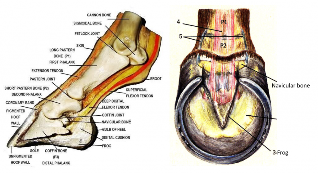
Horse Hoof Anatomy Labelled Teaching Chart Equine Foot lupon.gov.ph
Injecting the coffin joint with triamcinolone acetonide (TA) is a good way to deliver TA to the navicular bursa for horses with navicular syndrome. No studies have been done to show what level of TA is needed to control pain in the joint or bursa. Internal anatomy of the horse hoof. X-rays confirm needle placement in the navicular bursa.

Should I Give My Horse Joint Injections? Video Show Jumping Life
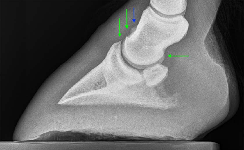
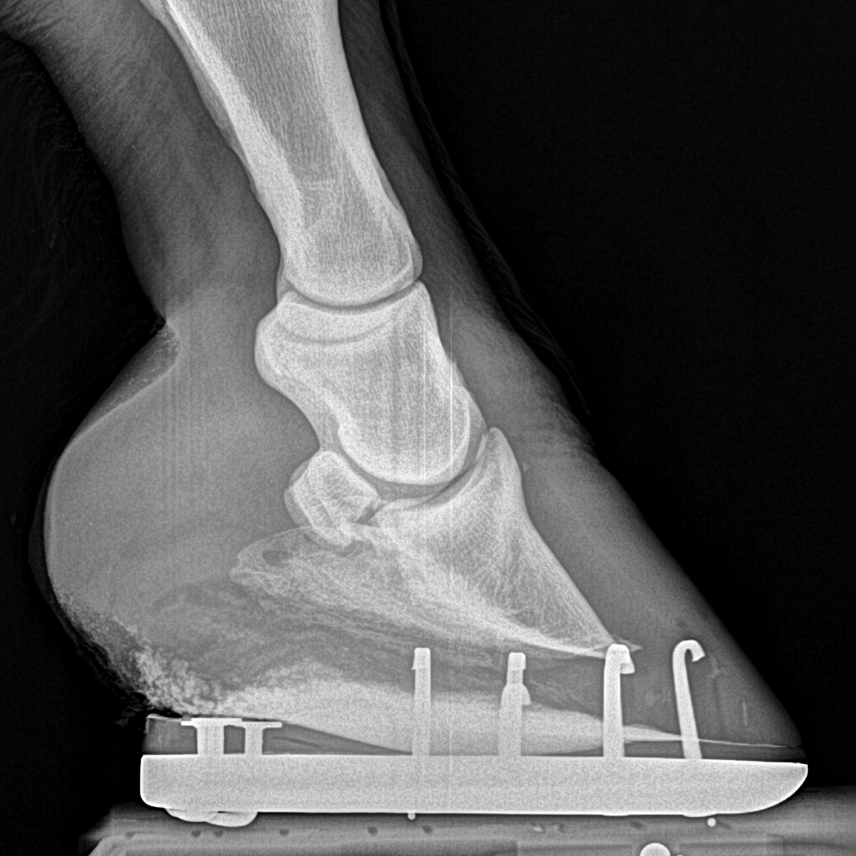
Blocking the coffin joint may also improve lameness in horses with navicular. Veterinarians have been able to show the deterioration of the navicular bone through radiograph footage. Figure 2 shows a radiograph of navicular. Using radiographs is highly debated when determining navicular disease because some horses may not show signs on a.
Treatment of lameness in the distal limb (coffin joint and pastern joint) of the horse
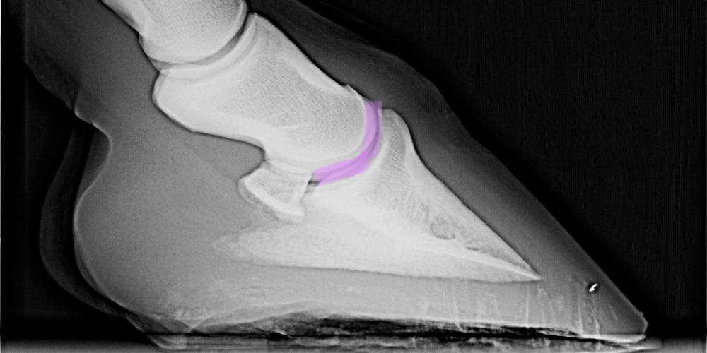
The coffin joint comprises the middle phalanx (short pastern bone), the distal phalanx (coffin or pedal bone) and the navicular bone. It has a voluminous joint capsule that extends upwards above.
What Are All The Bones That Make Up A Horse’s Hoof? Insider Horse Latest & Greatest Horse
The term ringbone describes the bony calcification that can develop on or around the pastern and/or coffin joints when arthritis develops in the joints. This can occur due to normal wear and tear or following injury and/or inflammation. This condition can occur in any foot (or even multiple feet). Ringbone can occur with arthritis of either the.
Piper’s Fractured Coffin Bone HorseDVM
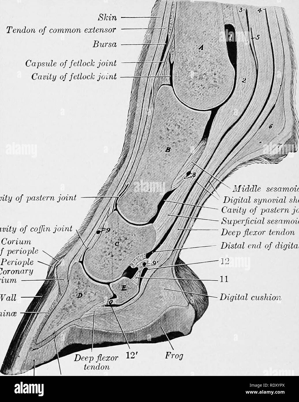
Coffin Joint. STEVE: The coffin joint is encased inside the hoof itself. It is a joint that exists between the third phalanx, or the coffin bone, and the short pastern bone. Another integral part is that the flexor tendon, which runs along and attaches on the base of the coffin bone — and has the navicular bone attached across it as it runs.
Common extensor tendon Black and White Stock Photos & Images Alamy
Laminitis Defined. Laminitis occurs when the lamellae (or laminae)—the Velcro-like tissues that suspend the coffin bone within the hoof capsule—become damaged and inflamed. In severe cases.

Coffin bone fracture in a horse.
In adult horses, coffin joint infections normally take a month or more to reveal their presence, usually in the form of a narrowed cartilage space. The delayed onset of radiographic indicators in adults is due largely to the loss of blood supply in the articular cartilage as the skeleton matures. Eventually, intraarticular bacteria enter the.

Michael Porter, Equine Veterinarian High Ring Bone!!
Additionally, Pluhar said bigger molecules are better for younger horses or high-motion joints like fetlocks, stifles and coffin joints. The bigger molecule blends are ones that cost the owner more money, but they offer a major benefit. "I like to use them in young horses to help make a horse's career longer.

Ligaments and tendons of the equine hoof. Horse anatomy, Horse health, Equine veterinary
The coffin joint—the joint between the coffin bone, the small pastern bone above it and the navicular bone tucked in the back—is at the core. When the horse loads the foot, his weight comes down through the small pastern bone and disperses through the navicular bone to the heel and through the coffin bone to the toe.
- Properties For Sale Broughty Ferry
- Local Life Has A Bad Start
- Fiat Ducato Fuse Box Diagram
- Paul Graham Estate Agents Sutton
- Black Friday Electric Scooter Deals
- The 1975 Live With The Bbc Philharmonic Orchestra Vinyl
- Toot Toot Monster Truck Rally
- Tga Breeze S4 With Canopy
- Big Bad Wolf Slot Game
- Is Bewdley Bridge Open Today